|
ABOUT US
The CCSVI-Center Frankfurt/ Germany was established in 2010 , inspired by Prof . Zamboni's effort with regards to the prospective vascular origin of multiple sclerosis and the demand by MS-patients for therapy .
 Due to the creativity of CCSVI , with handful of scientific studies being accessible to determine extended results , our principal objective at the CCSVI-Center Frankfurt is articulated by "primum nil nocere" – staying away from problems for patient as our major focus . Due to the creativity of CCSVI , with handful of scientific studies being accessible to determine extended results , our principal objective at the CCSVI-Center Frankfurt is articulated by "primum nil nocere" – staying away from problems for patient as our major focus .
To accomplish this milestone we have created partnership between professionals in the vast field of diagnostic imaging and vascular intervention who each are associated into a globally oriented system of professionals . This promises the maximum amount of competence in the judgment as well as treating CCSVI , the prevention of harmful side-effects as well as problems and the engagement in the speedy exchange of professional understanding .
WHAT IS CCSVI?
The close spatial partnership between demyelization plaques and venous vessels on which CCSVI based , was postulated before 1980s as as well the one-way link between venous outflows ailments in the brain and the improvement back marks of MS .
 Multiple sclerosis ( MS ) has been regarded as for quite some time to be an autoimmune ailment , but current investigation from Italy draws your attention to not an impossible different reason . In accordance with the theory of Prof . Paolo Zamboni , a vascular surgeon from Ferrara , narrowings ( stenoses ) in jugular veins ( neck ) or the azygos veins returning the blood from the head to the heart , could be accountable for the development and the symptoms of MS ( Multiple sclerosis ) . Multiple sclerosis ( MS ) has been regarded as for quite some time to be an autoimmune ailment , but current investigation from Italy draws your attention to not an impossible different reason . In accordance with the theory of Prof . Paolo Zamboni , a vascular surgeon from Ferrara , narrowings ( stenoses ) in jugular veins ( neck ) or the azygos veins returning the blood from the head to the heart , could be accountable for the development and the symptoms of MS ( Multiple sclerosis ) .
The blood vessels constriction puts a stop to blood running instantly enough ( so-called reflux , where the blood vessels would not function effectively and the blood flows into the brain once more ) , with the outcome that iron is laid on in the brain ( it could be seemed as white plaques visible on the MRI scan ) causing autoimmune swellingdamaging the protective myelin nerve coating in the spinal cord and in the brain .
This condition has become known as "CCSVI”: chronic cerebro-spinal venous insufficiency.
Widening of the stenotic (narrowed) veins increases the blood flow through them, thus, relieving the back-pressure and eliminating the inflammation, which is causing the damage to the brain, the spinal cord and protective myelin.
IDENTIFYING THE PROBLEM
CCSVI investigation and diagnosis at CCSVI-Center (Frankfurt/Germany):
MRV-Screening – CCSVI Diagnostics following a modified and advanced Haacke Protocol.
In order to diagnose the CCSVI, CCSVI-Center in Frankfurt uses non-invasive or mini-invasive CCSVI investigations methods. Our experience and our collaboration with the vendors of medical equipment and medical scientists around the world helped us to improve the CCSVI Diagnosis protocols, in order to provide reliable and safe diagnostic for our patients.
CCSVI DIAGNOSTIC:
Identification of CCSVI according to Zamboni is based on three methods:
Ultrasound examination (only head and neck)
-
The limitation of ultrasound is that it cannot assess the intrathoracic and abdominal venous system.
But it provides real-time information about blood flow dynamics, which is necessary to assess reflux in the lower internal jugular vein segment, that is important for the blood flow from the brain to the heart.
-
MR-Venography (head, neck, thorax and abdomen – Haacke protocol)
Improved Haacke Protocol by CCSVI-Center in Frankfurt
 |
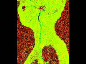
MR venography showing with color coding demonstrating focal stenosis of the upper internal jugular veins (IJV). |
|
We have implemented the following improvements for the Haacke protocol:
-
Combination of fast non breath-hold mr-venography (MRV) followed by slower breath-hold MRV for the lower neck and upper thoracic venous system
-
Flow-visualization along the internal jugular veins (IJV)
-
Flow visualization of the azygos vein-superior vena cava junction
-
Improved multi breath-hold single-volume equilibrium MRV of the thoracic and abdomino-pelvic venous system Examples of the respective improvements are show below.
The Haacke MR-protocol has been established as the standard for MR-venography and flow measurements in the work-up of CCSVI. It has, however, several drawbacks. Since MR-venography is carried out without breath holding and without cardiac gating, the azygos vein, that drains the blood from the spinal cord, cannot be depicted with sufficient accuracy on MRI
|
-
Catheter venography under fluoroscopy
Catheter venography or venogram, as a specialized form of angiography, is a medical imaging technique used to image the inside of blood vessels. This is traditionally done by injecting a contrast agent into the blood vessel and imaging with an X-Ray based technology such as fluoroscopy.
During catheter venography, a long, thin, flexible plastic tube (catheter) is inserted into the body, usually through a vein in the groin. The catheter is then threaded through the vascular system to the area requiring examination. A contrast dye is injected through the catheter and a rapid series of x-rays is taken, offering a detailed look at the specific blood vessels under the question.
 |
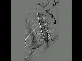
Catheter Venography showing the mild midlevel stenosis
|
THE COSTS FOR CCSVI INVESTIGATION
The costs for CCSVI investigations, including coordination, mediation and English language support depend on the art of the necessary investigation, additional packages and start from € 1050 to € 2500.
MEASURING RESULTS
Safety of the Treatment
Over the past 18 months we have successfully treated well over 400 patients, without complications.
Results of CCSVI and Multiple Sclerosis liberation Treatment
|
|
General Improvements |
Improvement oft the motoric system /
sensory system |
Treatment response in
fatigue |
complications |
|
strong |
60/100 |
40/100 |
35/100 |
0/100 |
|
moderate |
33/100 |
50/100 |
39/100 |
2*/100 |
|
Not any |
7/100 |
10/100 |
26/100 |
98/100 |
More than 90 % of our patients have an improvement after the treatment.

|


 Due to the creativity of CCSVI , with handful of scientific studies being accessible to determine extended results , our principal objective at the CCSVI-Center Frankfurt is articulated by "primum nil nocere" – staying away from problems for patient as our major focus .
 Multiple sclerosis ( MS ) has been regarded as for quite some time to be an autoimmune ailment , but current investigation from Italy draws your attention to not an impossible different reason . In accordance with the theory of Prof . Paolo Zamboni , a vascular surgeon from Ferrara , narrowings ( stenoses ) in jugular veins ( neck ) or the azygos veins returning the blood from the head to the heart , could be accountable for the development and the symptoms of MS ( Multiple sclerosis ) .

